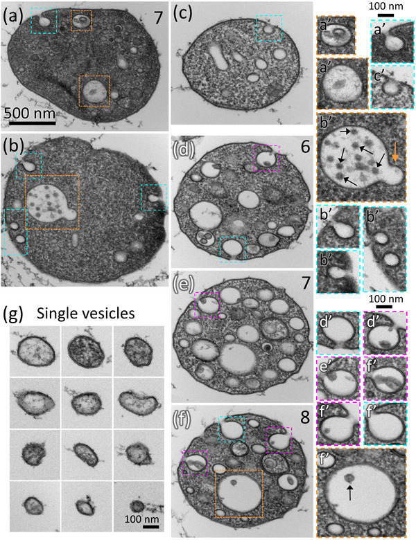
FIGURE 3.

MCMVs contain MVBs that fuse with the MCMV membrane to release exosomes. MCMVs containing organelles that are structurally similar or identical to MVBs are boxed orange in (a), (b), and (f) and shown to the right enlarged in orange boxes, (a’), (b’), and (f’). The MVB in (b’) appears to undergo an outward budding remodelling step (orange arrow). Black arrows indicate ILVs in (b’) and (f’). (a‐c) MCMVs with one or more omega‐figures on their limiting membrane that do not contain ILVs (boxed turquoise and enlarged to the right in turquoise boxes (a’), (b’), and (c’). (d‐f) Three serial sections through an MCMV (section number indicated in upper right corner) in which omega figures occur in each section. Some omega figures contain ILVs (magenta boxes, enlarged to the right in (d’), (e’), and (f’)) and some do not contain ILVs (turquoise boxes, enlarged to the right in (d’) and (f’)). (g) Single‐membrane vesicles located on the coverslip surface that are structurally like the inner vesicles of MCMVs. Scalebar in (a) applies to (a‐f). Supplemental Figure 6 and Movie 6 show serial sections of the MCMV shown in (a). Supplemental Figure 7 and Movie 7 shows additional serial sections of the MCMV shown in (d‐f).
