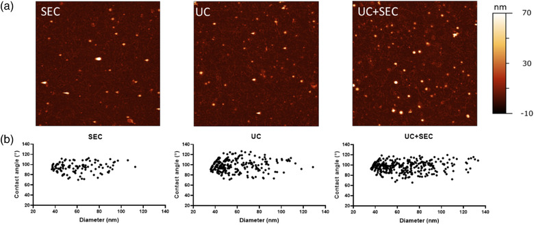
FIGURE 2.

Vesicle‐like structures are identified in all three preparations. (a) Representative AFM images of Ascaris suum EVs separated with either size exclusion chromatography (SEC), ultracentrifugation (UC), or a combination of those (UC+SEC). All AFM images are 5 × 5 μm. (b) Contact angle and equivalent diameter of Ascaris suum EVs obtained from AFM presented in scatterplots for each EV separation method
