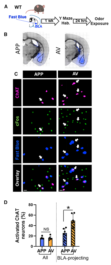
Figure 7. Although both APP and AV VP cholinergic neurons project to the BLA, the predominant BLA input from the VP stems from AV encoding VP cholinergic neurons.

(A) Workflow to retrogradely label BLA-projecting VP cholinergic neurons.
(B) Representative images in mice injected with fast blue in the BLA (in blue) and overlaid on a bright-field image.
(C) Representative images from the VP in a mouse exposed to the APP odor (left column) and AV odor (right column). First row, ChAT; second row, cFos; third row, Fast Blue; bottom row, overlay of all channels. Scale bars, 20 μm.
(D) AV responsive VP cholinergic neurons (n = 6 mice, all female) constitute a significantly greater percentage of BLA-projecting VP cholinergic neurons than APP responsive VP cholinergic neurons (n = 6 mice; 4 male and 2 female).
*p < 0.05. Error bars represent mean ± SEM.
