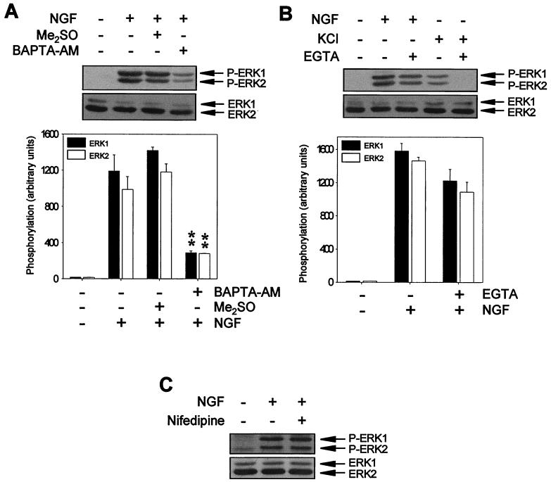
FIG. 1.
Ca2+ chelators block NGF-induced ERK phosphorylation. (A) PC12 cells were pretreated (+) or not pretreated (−) for 1 h with 50 μM BAPTA-AM or with vehicle (Me2SO) and then stimulated (+) or not stimulated (−) for 5 min with NGF. (B) PC12 cells were stimulated (+) or not stimulated (−) for 5 min with NGF or KCl in the presence (+) or in the absence (−) of 5 mM EGTA. (C) PC12 cells were pretreated (+) or not pretreated (−) for 1 h with 5 μM nifedipine and then stimulated (+) or not stimulated (−) for 5 min with NGF. After treatments, cells were lysed, and protein extracts were analyzed by Western blotting with an anti-phospho-ERK antibody (upper panels) and stripped and reprobed with an anti-pan-ERK antibody (lower panels) as a control for the protein content per lane. Graphs in panels A and B show the average ERK phosphorylation from three independent experiments. ∗∗, P value of <0.01, as determined by Student's t test. Arrows labeled P-ERK1 and P-ERK2 or ERK1 and ERK2 indicate the positions of phosphorylated and total ERK1 and ERK2 proteins, respectively.

