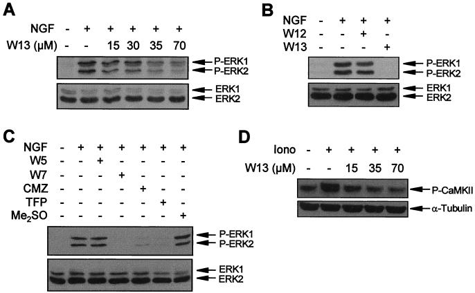
FIG. 2.
CaM inhibitors block NGF-induced ERK phosphorylation. (A) PC12 cells were pretreated or not pretreated (−) for 1 h with the indicated concentrations of the CaM inhibitor W13 and then stimulated (+) or not stimulated (−) for 5 min with NGF. (B) PC12 cells were pretreated (+) or not pretreated (−) for 1 h with W12 or W13 and then stimulated (+) or not stimulated (−) for 5 min with NGF. (C) PC12 cells were pretreated (+) or not pretreated (−) for 1 h with 100 μM W5 or W7, 25 μM calmidazolium (CMZ), 50 μM trifluoperazine (TFP), or vehicle (Me2SO) and then stimulated (+) or not stimulated (−) for 5 min with NGF. After treatments, cells were lysed and ERK phosphorylation was analyzed as described in the legend to Fig. 1. (D) PC12 cells were pretreated (+) or not pretreated (−) for 1 h with the indicated concentrations of CaM inhibitor W13 and then stimulated (+) or not stimulated (−) for 30 s with ionomycin (Iono). Lysates were probed with an antibody to phosphorylated CaMKII T286 (upper panel) and reprobed with an anti–α-tubulin antibody to assess comparable loading of lanes (lower panel).

