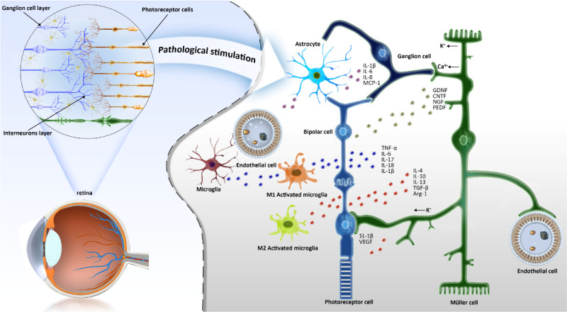
Figure 3.

Schematic diagram of the crosstalk between microglia and synaptic cells. In normal retinal tissue, ramified microglia are mainly distributed in the ganglion cell layer, inner plexus layer and outer plexus layer. When stimulated by pathology, microglia cells react quickly and change into an activated state. The interaction between microglia and Müller cells forms a trophic factor control system in the process of retinal degeneration. GDNF, glial cell line-derived neurotrophic factor; CTNF, ciliary neurotrophic factor; NGF, nerve growth factor; PEDF, pigment epithelium-derived factors; VEGF, vascular endothelial growth factor; TGF-β, transforming growth factor-β; MCP-1, Monocyte chemoattractant protein-1.
