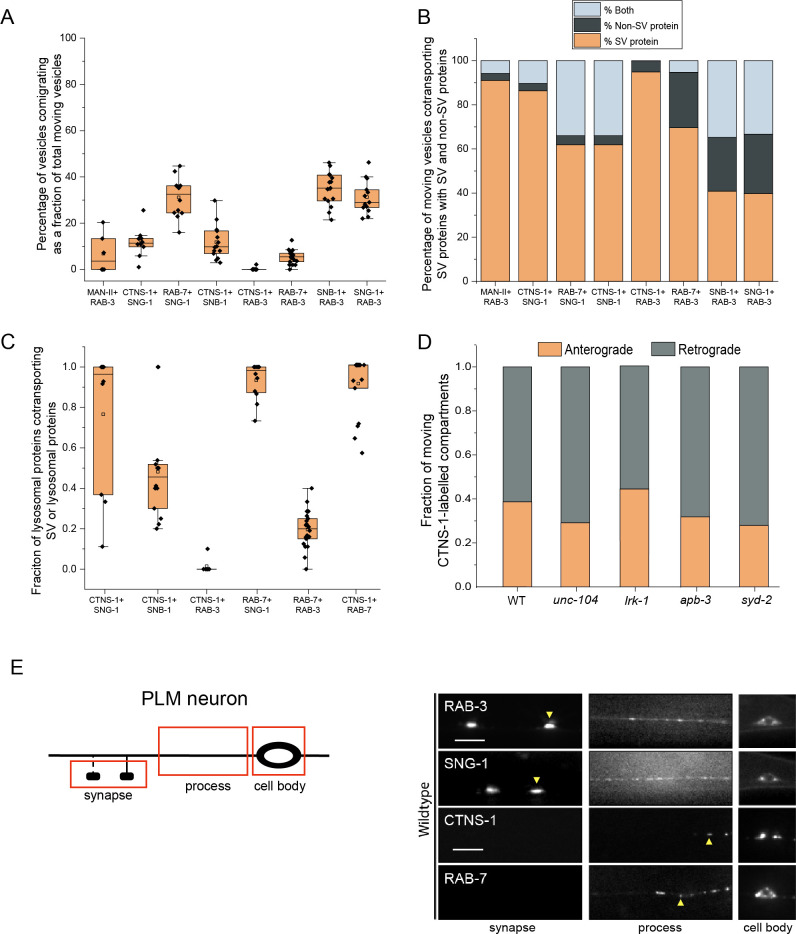
Fig 1. Synaptic vesicle proteins travel with lysosomal proteins in heterogenous carriers.
(A) Quantitation of percentage of total moving vesicles co-transporting different combinations of synaptic vesicle proteins and lysosomal proteins from kymograph analysis of dual color imaging. The number of animals per genotype (N) ≥ 10; number of vesicles analyzed (n) > 600. (B) Quantitation of percentage of total moving vesicles transporting different synaptic vesicle proteins or lysosomal proteins or both from kymograph analysis of dual color imaging. The number of animals per genotype (N) ≥ 10; number of vesicles analyzed (n) > 600. (C) Quantitation of fraction of various total moving lysosomal proteins co-transporting different synaptic vesicle proteins from kymograph analysis of dual color imaging. N ≥ 10; n > 100. (D) Quantitation of fraction of total moving CTNS-1-labeled compartments moving in the anterograde and retrograde direction in different mutants. N ≥ 9 per genotype; the number of CTNS-1-labeled compartments ≥ 20. (E) Schematic of the PLM neuron. Red boxes indicate the regions of imaging. The arrow shows the anterograde direction of vesicle motion. (F) GFP::RAB-3, SNG-1::GFP, CTNS-1::mCherry, and RAB-7::mScarlet in the cell body, process, and synapses of wildtype PLM neurons. Scale bar: 10 μm.

