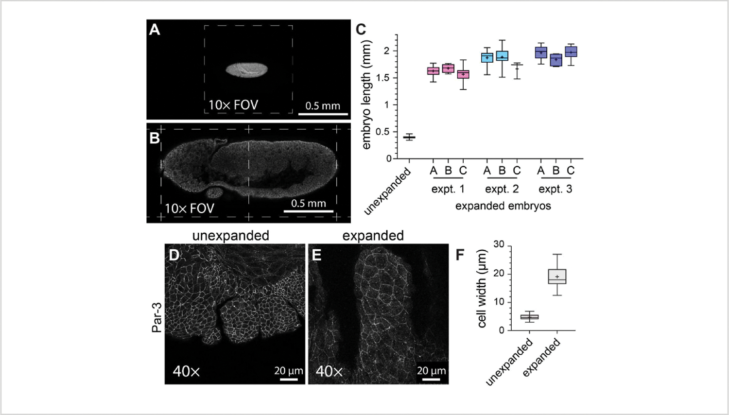
Figure 3: Four-fold expansion of Drosophila embryos.

(A) Unexpanded and (B) expanded Drosophila embryos imaged using a 10x objective (0.3 NA) at 1x zoom. Individual fields of view (FOV) are indicated with dashed lines. The embryos expressed a GFP-tagged version of myosin light chain and were stained with an anti-GFP antibody. (C) Quantification of embryo length (along the head-to-tail axis) in three hydrogels per experiment and from three separate ExM experiments compared with unexpanded controls. (D,E) Maxillary segments from (D) unexpanded and (E) expanded stage 11 Drosophila embryos imaged using a 40x objective (1.3 NA) at 1x zoom. The cell outlines (adherens junctions) were detected with an anti Par-3/Bazooka antibody (white). (F) Quantification of the cell width (long axis) from equivalent groups of cells from (D) and (E). The box plots in (C) and (F) show the 25th, 50th, and 75th percentile ranges; the whiskers indicate the minimum and maximum values; the “+” symbols indicate the mean.
