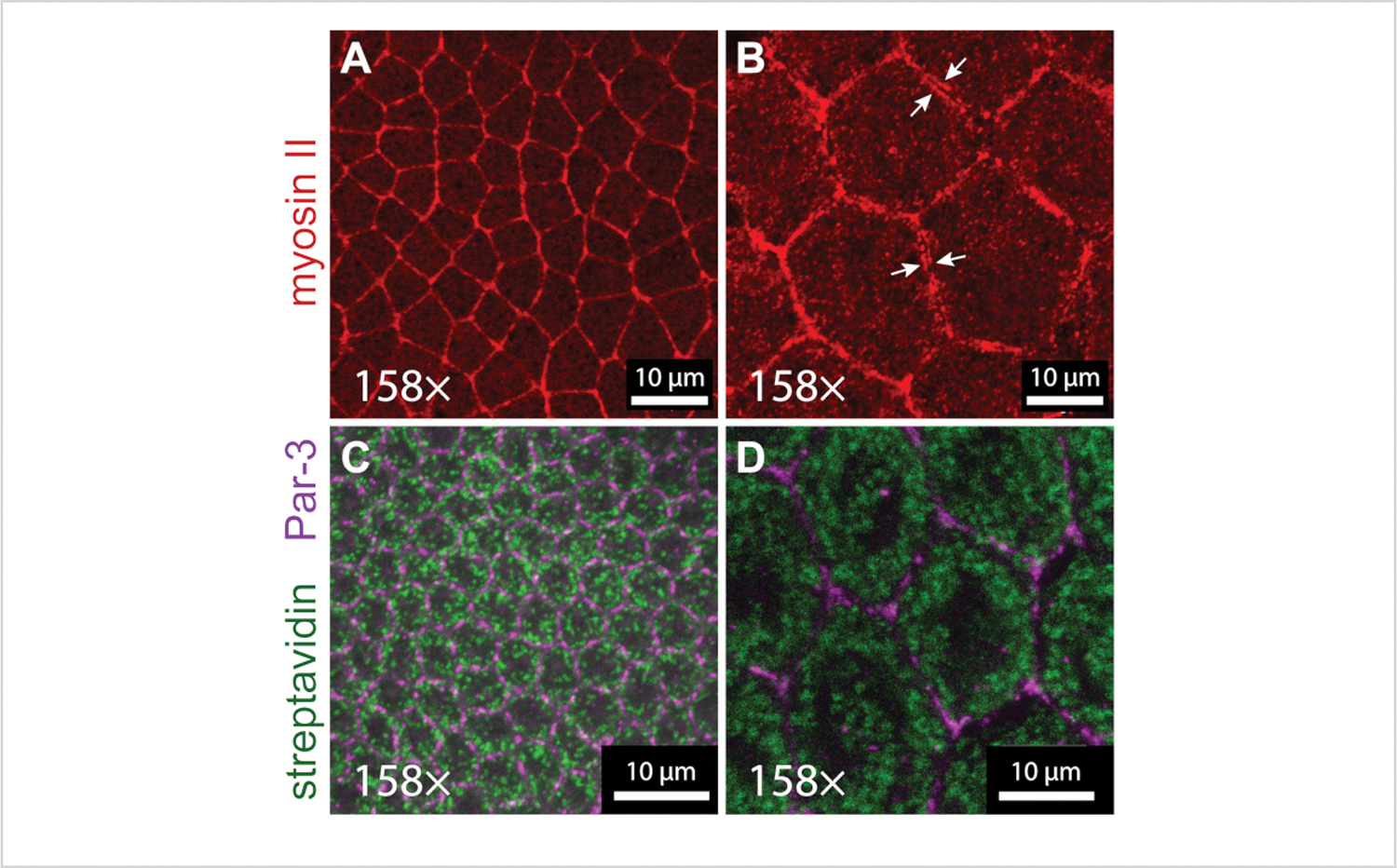
Figure 4: Details of actomyosin cytoskeleton and mitochondria revealed by expansion microscopy.

(A,B) Myosin II localization in neuroectoderm (germband) cells imaged with a 63x objective (1.4 NA) at 2.5x zoom in stage 7 (A) unexpanded and (B) expanded embryos. Myosin II was detected in the embryos expressing a transgenic GFP-tagged version of the myosin II regulatory light chain (sqh-GFP), which was detected with an anti-GFP antibody (red). Distinct pools of cortical myosin located in adjacent cells can be resolved in the expanded embryo (white arrows). (C,D) Mitochondrial networks in neuroectoderm cells imaged with a 63× objective (1.4 NA) at 2.5x zoom in stage 6 unexpanded (C) and expanded (D) embryos. The mitochondria were detected with streptavidin-Alexa 488 (green), and the cell outlines were detected with an anti-Par-3/Bazooka antibody (magenta). The experiments were performed with a laser-scanning confocal microscope.
