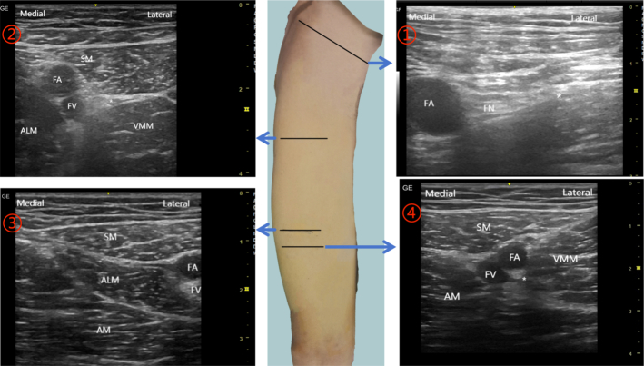
FIGURE 1.
Location of catheter insertion and corresponding ultrasound images. ①, The location of FNB, the ultrasonic probe was placed in the middle one-third of the transverse axis of the inguinal ligament.②, The location of FTB, the midpoint of the line connecting the apex of FT and ASIS. ③, The location of the apex of the FT (also the entrance of the AC). ④, The location of ACB, 3 cm below the AC entrance. ALM indicates adductor longus muscle; AM, adductor magnus muscle; FA, femoral artery; FN, femoral nerve; FV, femoral vein; SM, sartorius muscle, VMM, vastus medialis muscle; *target area.

