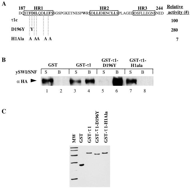
FIG. 3.
Binding of τ1 mutant proteins to the SWI-SNF complex. (A) Schematic representation showing the amino acid substitutions in the τ1 mutants used. Boxes indicate the locations of putative helical regions I, II, and III. Mean relative β-galactosidase activity (#) of τ1-core-LexA fusion proteins are shown as percentage of WT level (taken from reference 2). (B) Immunoblot showing coprecipitation of purified GST-τ1 mutant proteins with the SWI-SNF complex, which was detected by using an antibody raised against the HA-tagged SWI2 subunit. (C) Coomassie blue staining of GST-τ1 mutant proteins used in a pull down assay with the SWI-SNF complex.

