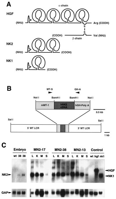
FIG. 1.
Structure and expression of NK2. (A) Schematic comparison of HGF/SF (designated as HGF in this and all other figures) and its natural splice variants NK2 and NK1. Each isoform contains a single so-called N domain at the amino terminus, and either four, two, or one kringle domain, as shown. However, only HGF/SF is processed into two chains, the β chain containing an enzymatically inactive serine protease domain. (B) The SalI-SalI NK2 transgene construct contained the human NK2 cDNA, the mouse MT gene promoter (mMT-1) and 5′ and 3′ flanking sequences (MT LCR), and the hGH poly(A) signal. Mice harboring the NK2 transgene were identified by PCR using primers MT-S and GH-A, as indicated. (C) Analysis of NK2 transgene expression in mouse tissues by Northern blot hybridization. For embryonic expression (three-lane panel at left), wild-type (wt) embryos were harvested at E16.5, and transgenic embryos from line MN2-38 (38) were harvested at E14.5 (middle lane) and E16.5 (right lane). Adult (2-month-old) tissues from three independently generated lines, MN2-17, MN2-38, and MN2-13, were studied. Tissues analyzed included liver (L), kidney (K), skeletal muscle (M), and skin (S). The control lanes at far right show expression of HGF/SF sequences in livers of wild-type and HGF/SF and NK1 transgenic mice. Following hybridization with a human NK2 cDNA probe (top panels), the filter was stripped and rehybridized with a control GAP cDNA probe (bottom panels).

