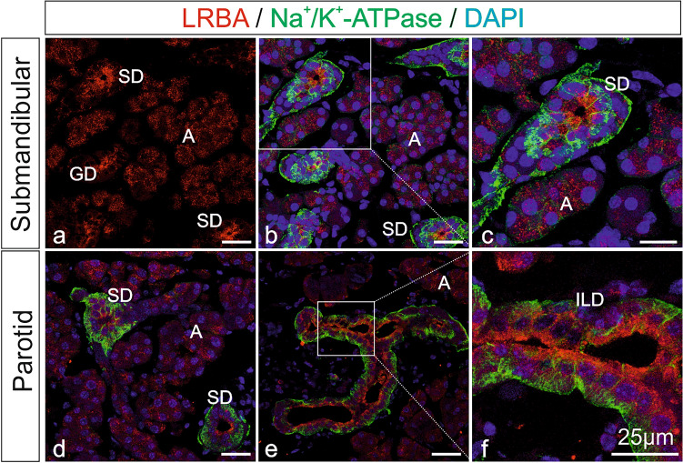
Figure 4.
LRBA immunofluorescence in salivary glands. Double-immunofluorescence confocal microscopy on mouse submandibular and parotid gland for LRBA (red) and for Na+/K+-ATPase (green) as a marker of basolateral plasma membranes. Nuclei were stained with DAPI. (c,f) Represent higher magnification images from the white-boxed areas in (b,e), respectively. LRBA immunofluorescence concentrated toward the subapical and apical cytoplasm of acinar and duct cells, and was abolished in KO control tissue (Supplementary Fig. S1b). A acinar cells, GD granular duct, ILD intralobular duct, SD striated duct. All scale bars: 25 µm.

