Figure 6.
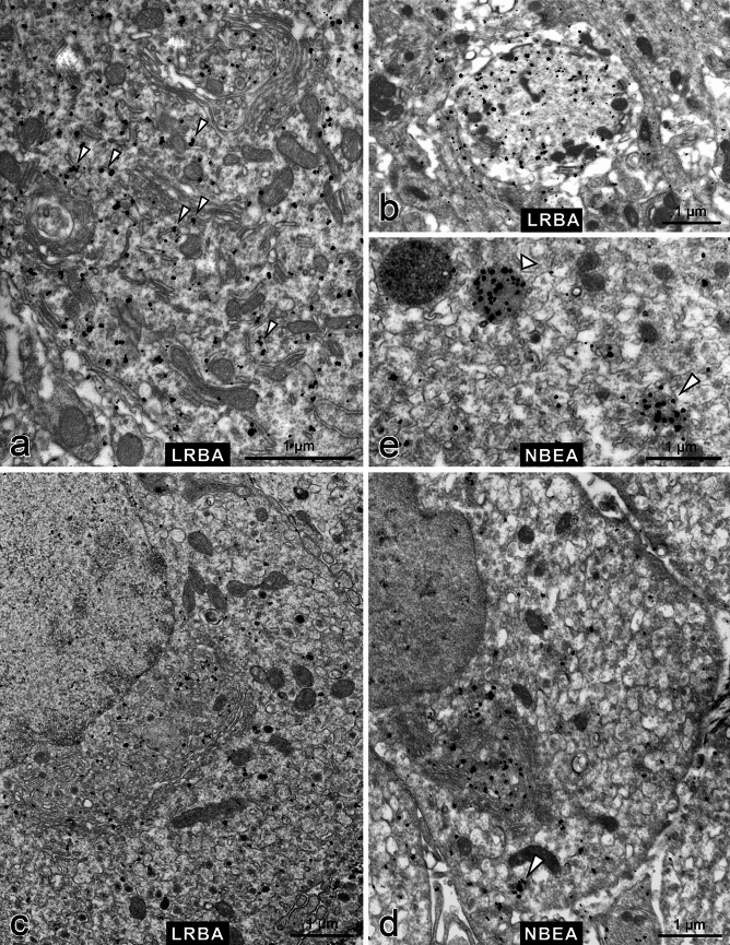
Immuno electron microscopy demonstrates differential subcellular distributions of LRBA and NBEA. Pre-embedding immunolabelling with anti-LRBA (a–c) or anti-NBEA (d,e) was followed by nanogold-conjugated secondary antibody and silver-enhancement. (a) Purkinje cell cytoplasm with two Golgi complexes (top and center) and occasional association of LRBA immunolabel with endomembranes (arrowheads). See also Supplementary Fig. S2c. (b) Cross-sectioned Purkinje cell dendrite (LRBA-positive) surrounded by the largely LRBA-negative neuropil of the molecular layer of the cerebellum. (c) Adrenal chromaffin cell with a typical, convoluted paranuclear Golgi complex; LRBA immunolabel is scattered throughout the cytoplasm, slightly enriched near the concave face of the Golgi complex. See also Supplementary Fig. S2d. (d) Adrenal chromaffin cell; NBEA immunolabel is strikingly enriched in the concave space of the Golgi complex. See also Supplementary Fig. S2e. (e) Close-up of a chromaffin cell shows NBEA immunolabel decorating the lumen of a lysosome-like organelle (broad arrowhead) whereas another putative lysosome to the left is NBEA-negative; and an aggregate of small vesicles and diffuse material (slim arrowhead). Such an aggregate is also pointed out by an arrowhead in part (d). See also Supplementary Fig. S2f.

