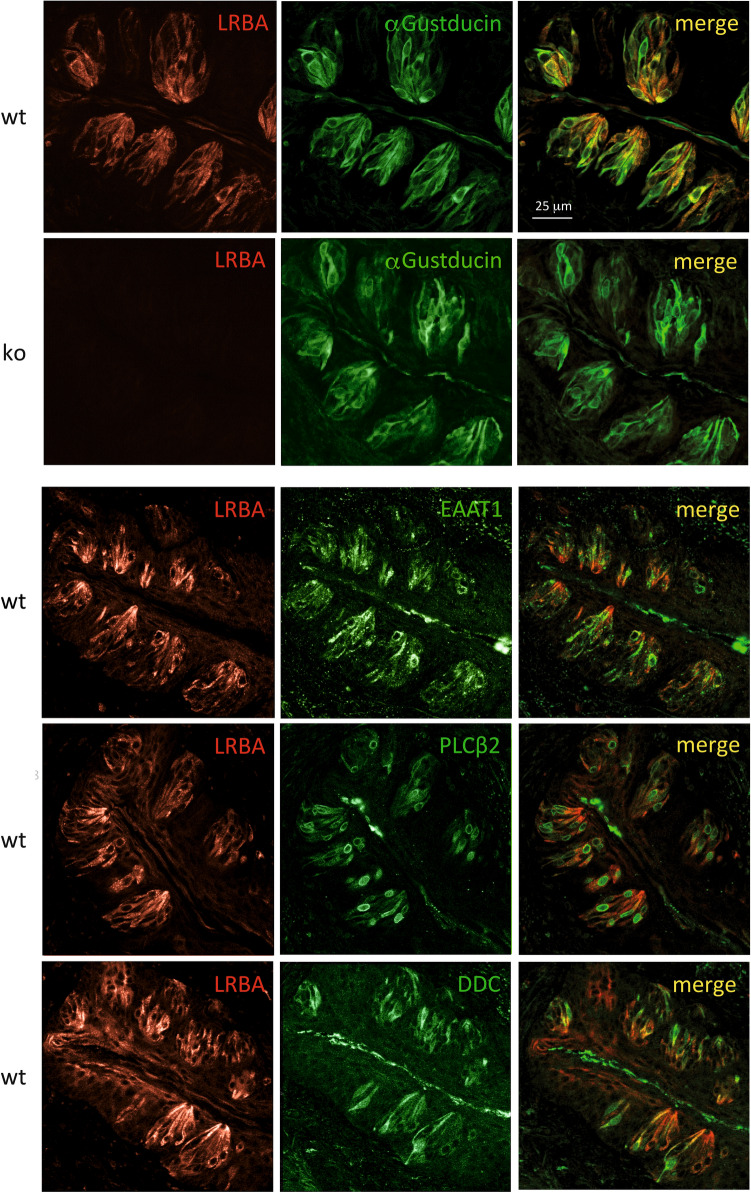
Figure 7.
LRBA immunofluorescence in type I–III taste cells of vallate papilla taste buds. LRBA-IF-positive cells (left column) display high degrees of overlap with cells IF-positive for α-Gustducin, EAAT1 (type I cell marker), PLCβ2 (type II cell marker) and DDC (type III cell marker). In taste buds from LRBA-KO mice, LRBA-IF was not detectable, while the staining patterns of α-Gustducin (second row from top) or the other markers (not shown) seemed unaffected by the LRBA-KO.

