Figure 3.
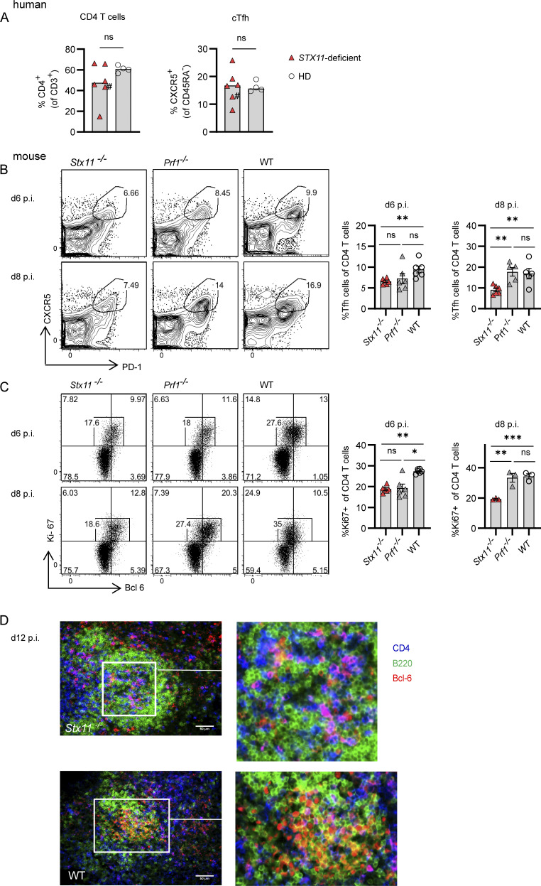
Reduced Tfh numbers in Stx11−/− mice during LCMV infection. Human: (A) Percentages of CD4 T cells and Tfh (CXCR5+ CD45RA−CD4+) cells in frozen PBMCs samples from FHL-4 patients (red triangle) and healthy controls (open circle); #, patient under treatment. Mouse: (B–D) Stx11−/−, Prf1−/−, and WT mice infected with 200 PFU LCMV-WE i.v. (B and C) (B) Frequency of CXCR5+PD-1+ Tfh cells Stx11−/−(n = 6), Prf1−/− (n = 5–6), and WT (C57BL/6N n = 5–6) from n = 2 independent experiments and (C) proliferation by Ki-67 expression of Bcl-6+ Tfh cells from the spleen of Stx11−/−(n = 3 [d8]; n = 6 [d6]), Prf1−/− (n = 3 [d8]; n = 6 [d6]) and WT (C57BL/6N n = 3 [d8]; n = 6 [d6]) were analyzed on day 6 (n = 2 independent experiments) and d8 (1 experiment) p.i. (D) Representative immunofluoresent spleen sections: B220 (green), CD4 (blue), and BCL-6 (red) d12 p.i. with LCMV; WT (C57BL/6N) n = 6 of two independent experiments and Stx11−/− n = 3 of two experiments. Scale bar 50 µm. (A–D) Mean and ± SEMs of Mann–Whitney U test are shown; *P < 0.05, **P < 0.01, *** P < 0.001, ns indicates not significant.

