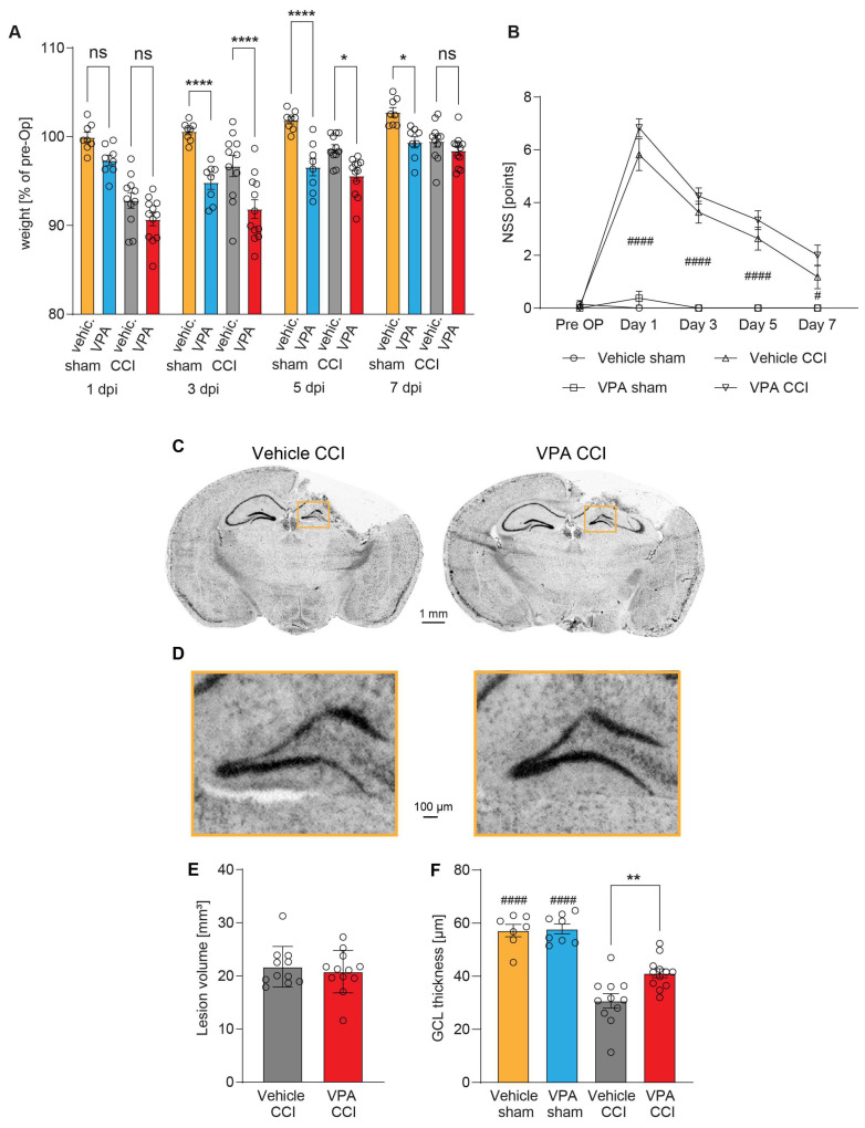
Figure 3.
Administration of VPA does not influence acute neurological deficits or brain lesion size at 7 dpi but attenuates structural damage in the hippocampus. (A) Body weight time course at post-traumatic day 1, 3, 5 and 7 in % of pre-surgery body weight [g], ns = not significantly different. (B) Neurological severity score (NSS) day 1, 3, 5 and 7. Sample size NSS/body weight: vehicle-controlled cortical impact (CCI): n = 11, VPA CCI: n = 12, vehicle sham: n = 8, VPA sham: n = 8. * p < 0.05, **** p < 0.0001 significantly different as indicated (CCI vs. sham animals). (C) Representative images of cresyl violet stained brain sections at 7 dpi from vehicle or VPA-treated mice. (D) Boxed regions from (C) shown in higher magnification with detail enlargement of the hippocampal granule cell layer (GCL) at 7 dpi in vehicle and VPA-treated mice. (E) Quantification of lesion volume and (F) GCL thickness (vehicle CCI: n = 11, VPA CCI: n = 12), * indicates significance levels between CCI groups and # between sham and corresponding CCI groups (** p < 0.01, #### p < 0.0001). All data points represent individual animals, and data are expressed as mean ± SEM, p values were calculated by two-way ANOVA with Holm–Šidák correction (A), Kruskal–Wallis test with Dunn’s correction (B), Mann–Whitney U Test (E) and one-way ANOVA with Holm–Šidák correction (F).

