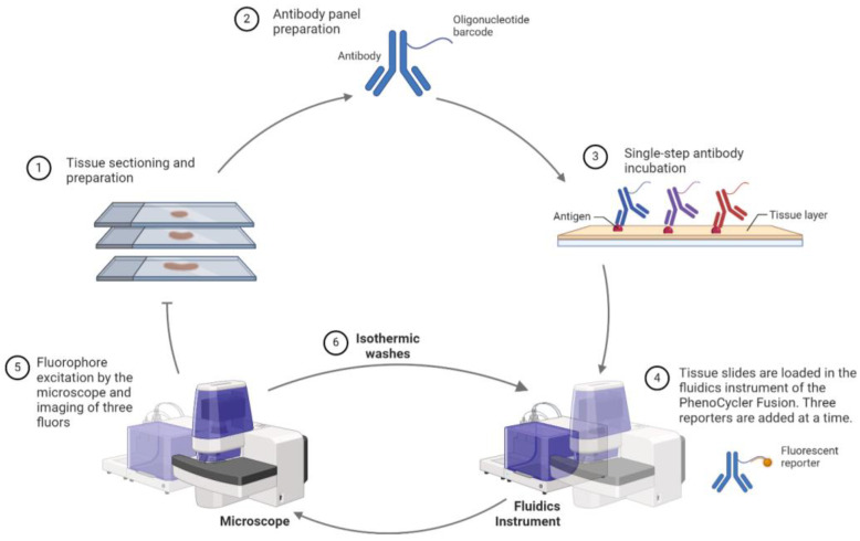
Figure 5.
Workflow of the Akoya PhenoCycler Fusion. FFPE tissue sections are stained with antibody panels linked to oligonucleotide barcodes. The PhenoCycler cycles through staining, imaging, and barcode removal, enabling visualization of a comprehensive set of markers. Following antibody incubation, fluorescent reporters bind to the complementary barcode on the antibody. Three reporters are added at a time, and subsequent isothermic washes allow the addition of the following set of reporters. Created with Biorender.com.

