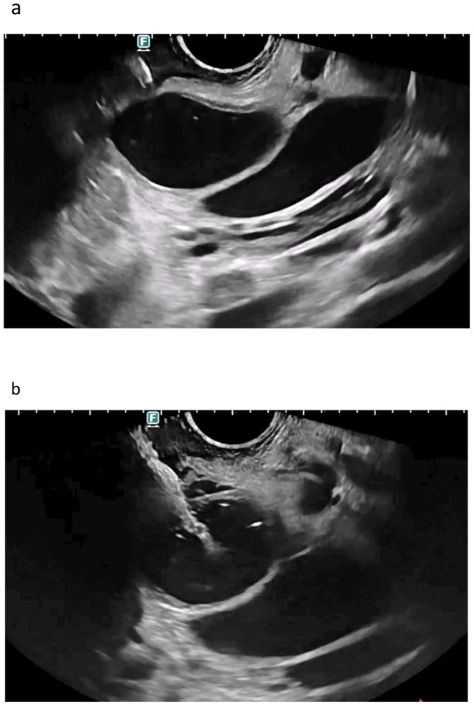
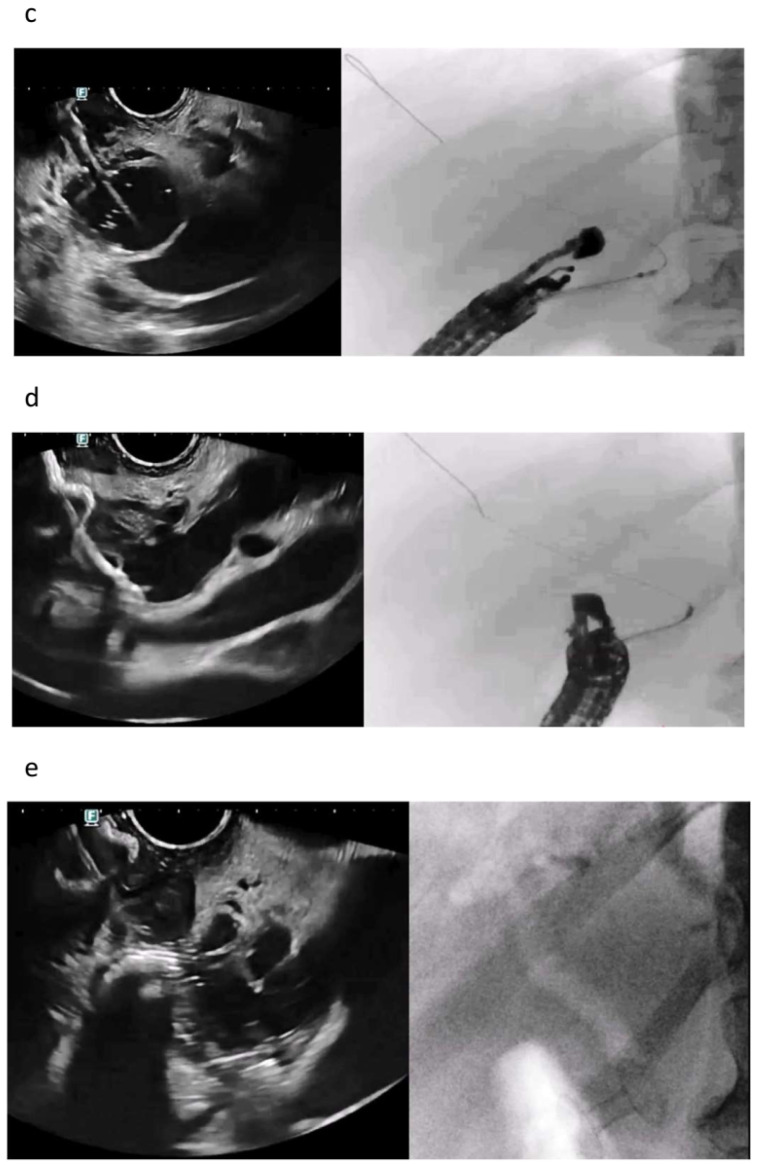
Figure 1.
EUS-CDS misdeployment. Anatomical delineation of the common bile duct next to the tip of the instrument (a). Tip of the electrocautery-enhanced lumen-apposing metal stent before deployment (a). Distal flange of the LAMS opened transmurally between the bile duct and the duodenal wall (b). Despite the flange being opened transmurally, the tip of the device was correctly inside the bile duct, and therefore, a guidewire was moved toward the hilum (c). The LAMS was recaptured and moved over the wire for a fluoroscopy-guided release, with the correct placement of the distal flange inside the bile duct (d). Final placement of the LAMS with the distal flange inside the bile duct and the proximal flange in the duodenum (left: endosonography; right: fluoroscopy confirming aerobilia through the LAMS (e).


