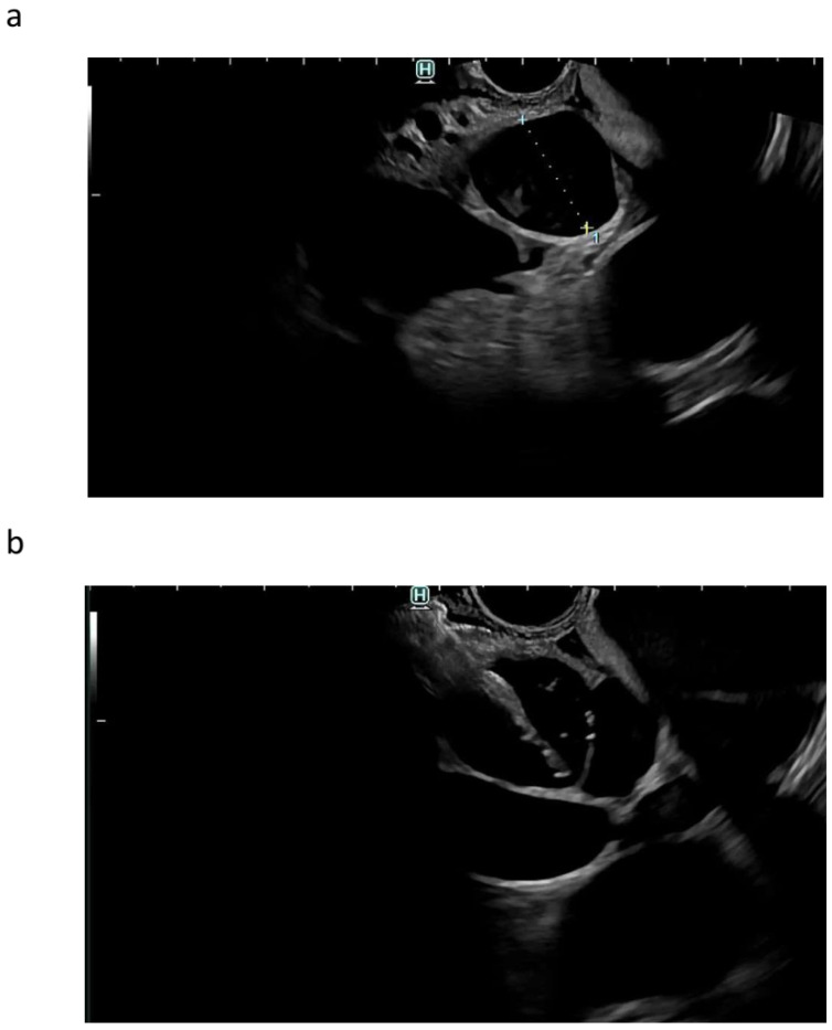
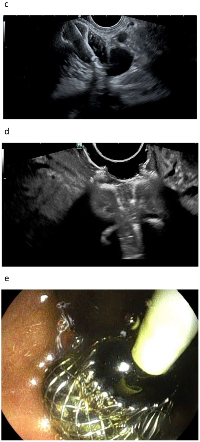
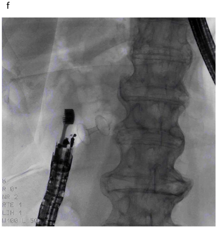
Figure 3.
EUS-guided choledochoduodenostomy. EUS-guided identification of the dilated common bile duct above the neoplasia from the bulb (a). EUS-guided penetration of the common bile duct with the electrocautery-enhanced lumen-apposing metal stent (b). Release of the distal flange inside the common bile duct, and traction in preparation for the intrachannel release of the proximal flange (c). EUS appearance of the released stent (air [CO2] flowing inside the stent) (d). Endoscopic appearance of the stent, draining bile (e). Radiologic appearance of the stent, with aerobilia depicting the biliary tree (f).


