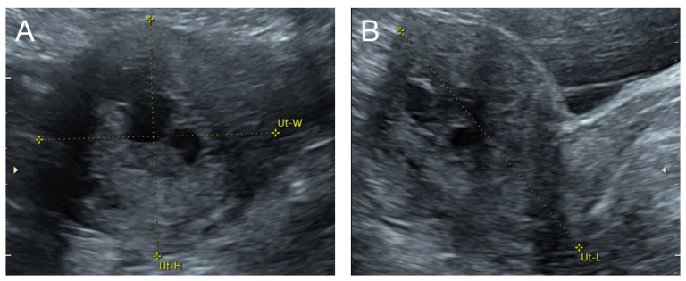
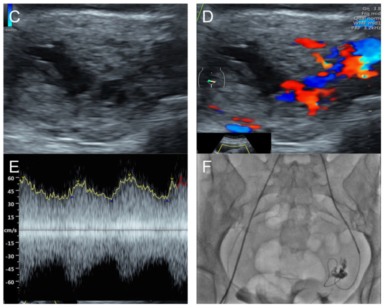
Figure 1.
(A–C) Transabdominal grayscale ultrasound: cross-sectional, sagittal, and oblique scans of the uterus showed heterogeneous soft tissue content, like pieces of conceptive product in the uterine cavity; ill-defined endometrial–myometrial interface; hypoechoic lacunae varying in size in the non-specific tissue content, mainly localized at the left anterior wall. (D) Color flow mapping with a relatively high pulse repetition frequency of 3.2 kHz (applied to the same image (C)) showed hyper-vascularized lesions in the myometrium; multidirectional flow, mainly localized at the left anterior wall; and some cystic spaces of no flow, indicating lysed blood in the cavity. (E) Spectral Doppler ultrasound showed a high peak systolic velocity (approximately 60 cm/s). The sonographic diagnosis was uterine AVM. The main differential diagnoses were incomplete abortion (conceptive products) and gestational trophoblastic disease. (F) CTA during uterine embolization revealed hypervascularity and tortuous arterial anatomy enhancing a dilated vascular pouch overlying the endometrium of the uterus with feeding via the bilateral uterine arteries and draining via the internal iliac veins, confirming uterine AVM; low blood content in the uterine cavity without evidence of active contrast extravasation.


