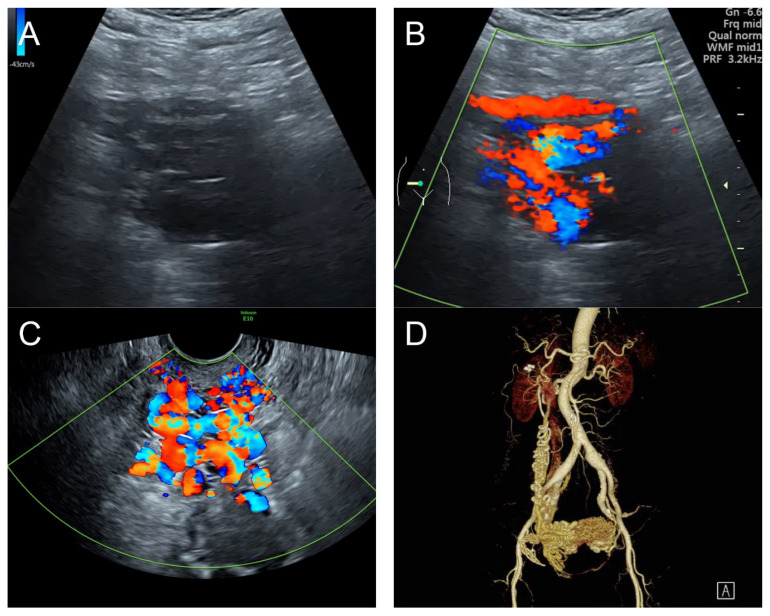
Figure 2.
(A) Transabdominal grayscale ultrasound: transverse scans of suprapubic area showed suboptimal-quality image of the uterus, which displayed ill-defined soft tissue mass, non-visualized endometrial lining, hypoechoic areas of the uterus. No normal architecture of the uterus could be demonstrated. (B) Color flow mapping (the same area of figure (A)) showed markedly vascularized uterus, involving throughout the uterus intense vascularity with a chaotic, multidirectional flow. (C) Transvaginal color Doppler ultrasound showed markedly vascularized uterus, involving throughout the uterus intense vascularity with a chaotic, multidirectional flow. (D) Abdominal CTA revealed innumerable tortuous dilated vessels in the pelvic cavity, along the entire uterine wall. The lesions were fed by multiple arterial feeders, which were displayed as tortuous dilated arteries of the bilateral uterine arteries, right ovarian artery, and right inferior epigastric artery. Multiple draining veins demonstrated early venous opacification, namely the bilateral internal iliac veins and right ovarian vein. The CTA findings confirmed uterine AVM.

