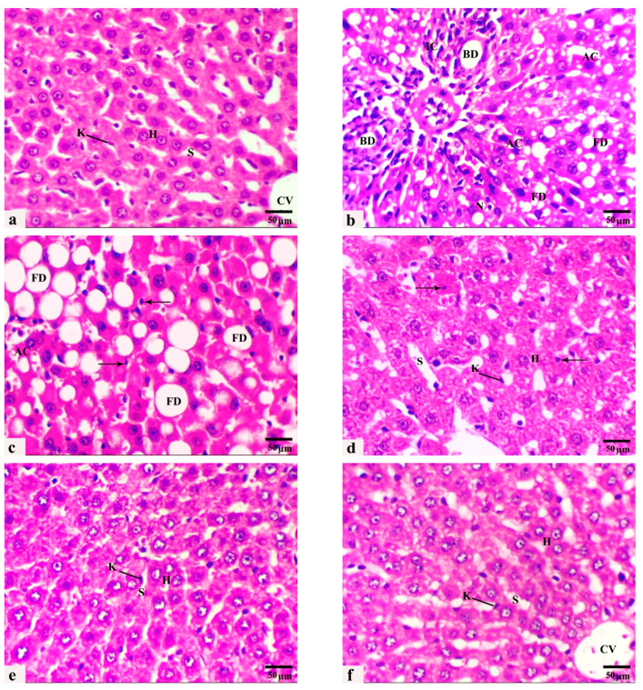
Figure 3.
(a–f): Photomicrographs obtained from H. and E. liver sections of (a) control rats displaying normal radial cords of hepatocytes (H), central vein (CV), sinusoid (S), and Kupffer cell (K). (b,c) HCD-fed rats showing microvesicular steatosis with inflammatory cell infiltration (IC), bile ductulus proliferation (BD), fatty degeneration of swollen hepatocytes with large coalesced vacuoles (FD), apoptotic cell (AC), necrotic area (N), and pyknotic nuclei (thin arrows). (d) rats that received HCD + L-carnitine showed a moderate improvement in hepatic architecture. Hepatocytes (H), dilated sinusoids (S), Kupffer cells (K), and pyknotic nuclei (thin arrows). (e) rats that received HCD + Ginkgo biloba displayed an obvious improvement in hepatic architecture, hepatocytes (H), dilated sinusoids (S), and a few activated Kupffer cells (K). (f) Rats that received HCD + LC + GB showed nearly normal hepatic strand architecture, central vein (CV), hepatocytes (H), sinusoids (S), and Kupffer cells (K). H. and E. stain, magnification ×40, scale bar 50 µm.

