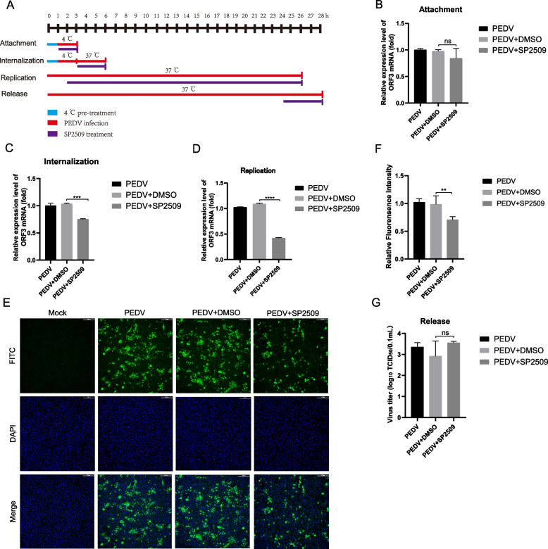
Fig. 4.
The antiviral effect of SP2509 on different infection steps of PEDV.The supernatant was replaced with SP2509 during the virus's attachment, invasion, replication, and release phases. A SP2509 treatment schemes. The blue bar represents 4℃ pretreatment, the red bars represent PEDV infection, the purple bars represent SP2509 treatment, and the vertical bars represent the cell collection. B Virus attachment assay. RT-qPCR detected PEDV-ORF3 mRNA level. C Virus internalization assay. PEDV-ORF3 mRNA level was detected by RT-qPCR. D Virus replication assay. PEDV-ORF3 mRNA level was detected by RT-qPCR. E IFA detection of SP2509 on PEDV replication phase. F IFA fluorescence quantification results. G Virus release assay. A virus in the supernatant was detected by TCID50. Data are expressed as three independent experiments. ns P > 0.05, * P < 0.05, ** P < 0.01, *** P < 0.001, **** P < 0.0001

