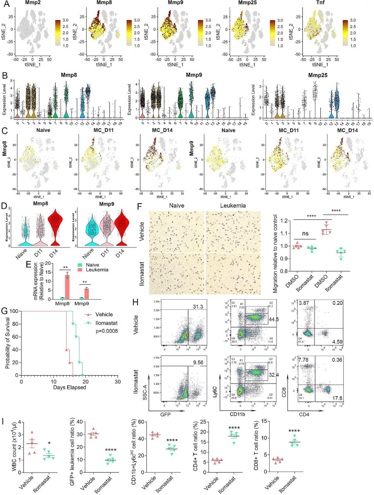
Fig. 6.
MMP pathway genes are involved in neutrophil mobilization and recruitment. Feature plots of gene expression of the Mmps in the TME of leukemic mice (A) show absence of expression of Mmp2 throughout and increased expression of Mmp8, 9 and 25 in different neutrophil populations, with predominant expression in the Ly6g + and Camk1d + neutrophils. In contrast, high-level Tnf expression is mostly restricted to the Il1b + neutrophils. The relative expression levels of Mmp8, 9 and 25 across the TME clusters is shown in the violin plots in (B). Feature plots for Mmp8 and 9 during leukemogenesis are shown in (C) and expression levels for Mmp8 and 9 during leukemia progression are shown in the violin plots in (D). Expression levels of Mmp8 and Mmp9 (N = 3) as determined by qPCR (E) shows significantly increased expression levels in neutrophils from the leukemic mice compared with naive mice. From in vitro migration assays, Cystal Violet staining (F, left) shows that neutrophils from leukemic mice show significantly higher cell motility compared with naïve neutrophils. In addition, while Ilomastat treatment does not affect isolated neutrophils from naive mice, neutrophils from leukemic mice show significantly decreased migration compared with DMSO-treated cells. The migration levels for each cohort (n = 5) is shown in (F, right). Kaplan-Meier analysis of survival in vivo shows that leukemic mice treated with Ilomastat have a significantly increased survival compared with vehicle control treated mice (G). Flow cytometric analysis (H) shows a reduction in GFP + leukemic cells in Ilomastat treated mice with a concomitant increase in CD4+/CD8 + T-cells and a decrease in CD11b + Ly6CInt PMN-MDSCs. Levels of these cells from all five mice in each cohort are shown in (I)

