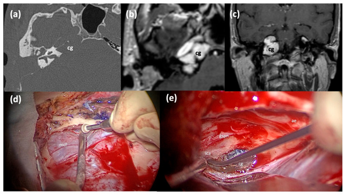
Figure 5.
Middle cranial fossa approach. (a) Axial CT scan of a right Type C PACG with erosion of the horizontal ICA bone walls. (b,c) Axial and Coronal T1-weighted MRI sections of the same PACG, showing the typical hyperintense signal. (d) Surgical step: a craniotomy has been performed and the dura of the middle cranial fossa is carefully elevated from the skull base. (e) Surgical step: the lesion is identified after the Kawase triangle drilling. cg: cholesterol granuloma; mcf, middle fossa dura; gspn, greater superficial petrosal nerve; ae, arcuate eminence; ica, internal carotid artery.

