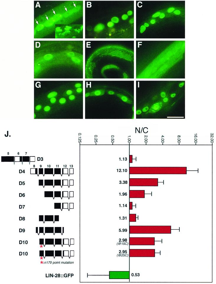
FIG. 4.
Nuclear localization efficiency of various truncated LIN-14::GFP proteins. (A to I) Hypodermal cells expressing LIN-14D3::GFP (A) LIN-14D4::GFP (B), LIN-14D5::GFP (C), LIN-14D6::GFP (D), LIN-14D7::GFP (E), LIN-14D8::GFP (F), LIN-14D9::GFP (G), LIN-14D10::GFP (at 15°C) (H), and LIN-14D10::GFP (at 25°C) (I). Animals were at developmental stages ranging from L1 to L3. (E and F) Unlocalized LIN-14D7::GFP and LIN-14D8::GFP appear as a uniform green fluorescence, while in all other panels except A; the green fluorescent spots are nuclei in which LIN-14::GFP is localized. Nucleoli are relatively free of LIN-14::GFP staining and appear as darker spots within the nuclei. The nuclear localization patterns of LIN-14D1::GFP and LIN-14D2::GFP (data not shown) are similar to that of LIN-14D4::GFP. (A) Lateral hypodermal seam cells (arrow) are filled with unlocalized LIN-14D3::GFP and show a higher level of GFP fluorescence than surrounding hypodermal cells, and the inset shows the unidentified GFP-containing inclusions that are frequently evident in animals expressing LIN-14D3::GFP. We have not determined whether the structures are extracellular or intracellular. The image was captured with relatively short exposures (1/30 or 1/60 s) so the actual level of GFP fluorescence is much higher than in the rest of panels, which were taken at exposures ranging from 1/4 to 2 s. (J) Quantitative assay of the nuclear localization efficiency of truncated LIN-14::GFP proteins. In the diagram on the left, black regions in exon boxes represent the amino acid sequences that are relatively well conserved among LIN-14 proteins from different nematode species (see Fig. 3). GFP sequences are not shown, and the intron spaces in D3 are not drawn in scale. N/C, nuclear-to-cytoplasmic ratio of GFP fluorescence plotted in a log scale (see Materials and Methods). LIN-28::GFP is a cytoplasmically localized protein (4) used here as a control. Bar, 5 μm.

