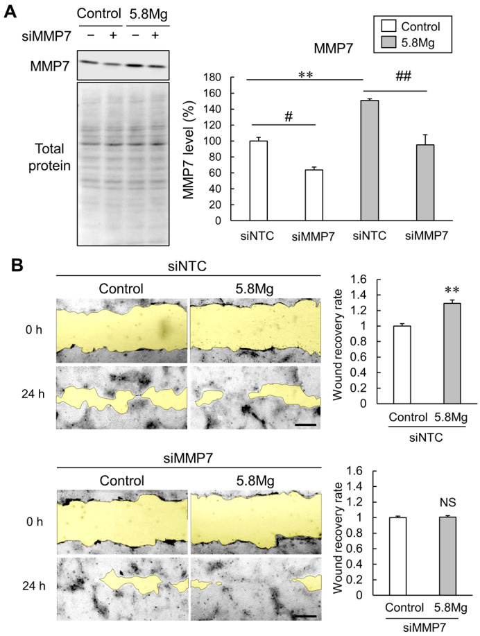
Figure 5.
Effect of MMP7 knockdown on Mg2+-induced cell migration promotion. (A,B) HaCaT cells transfected with 50 nM siRNA for a negative control (siNTC) and with MMP7 (siMMP7) were cultured for 48 h to knockdown the expression of MMP7. (A) HaCaT cells were cultured with DMEM containing 0.8 mM MgCl2 (Control) and 5.8 mM MgCl2 (5.8 Mg) for 24 h after MMP7 knockdown. After collecting the cells, MMP7 protein levels were analyzed using Western blotting. n = 4. ** p < 0.01, vs. siNTC control. ## p < 0.01, # p < 0.05 vs. siNTC. (B) The cells were cultured in 0.5% FBS medium supplemented with Control and 5.8 Mg conditions and scratched with a pipette tip after MMP7 knockdown. The typical images of the cells are presented. Yellow shows the wounded area. The scratched area is examined for the wound recovery rate (ratio to control) at 24 h. n = 4. NS p > 0.05 vs. Control.

