Figure 3.
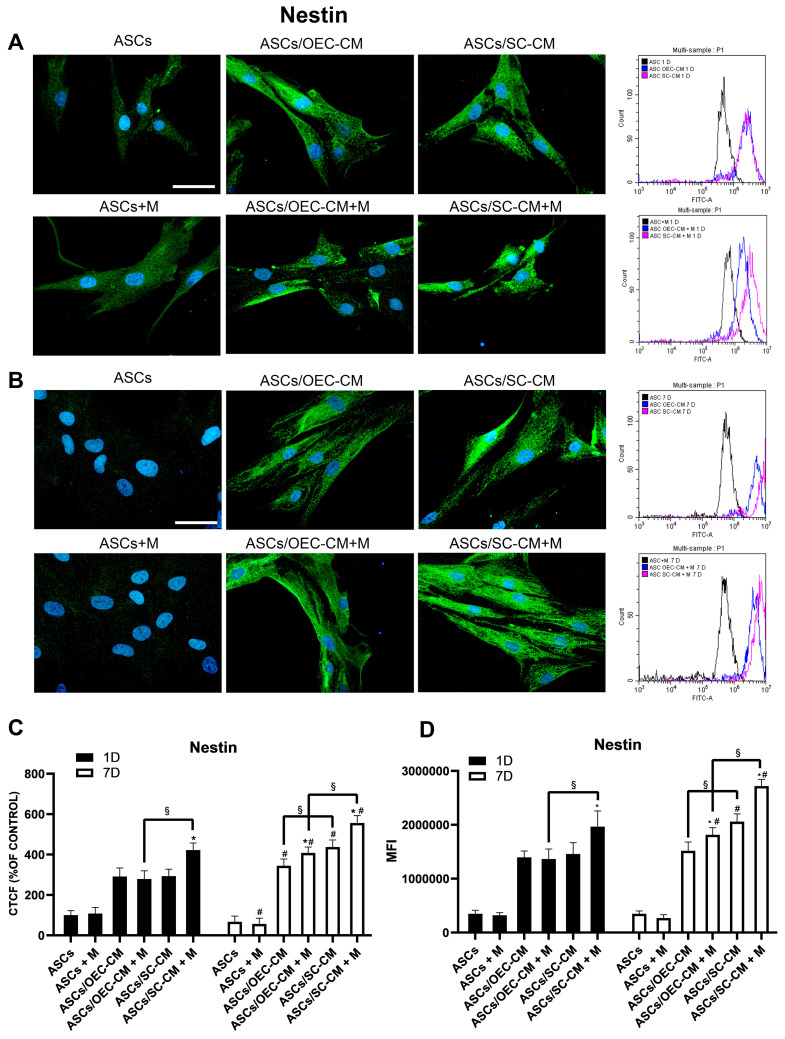
Nestin expression in ASC cultures. The different conditions were investigated by immunocytochemistry and flow cytometry on day 1 (A) and 7 (B) of culture. In each row, microphotographs are followed by flow cytometry histograms (fourth column) in the same conditions. Quantitative immunofluorescence data (ImageJ) and flow cytometry mean fluorescence intensity (MFI) are also reported in panels (C,D), respectively. A weak Nestin expression is detected when ASCs are grown in their basal medium, after both 1 and 7 days, regardless of the presence of melatonin (first column). A substantial increase is, however, observed when glial CM are used (second and third columns). Blue fluorescence counterstaining indicates cell nuclei. Scale bars: 50 µm. Quantitative data reported in the histograms (panels C,D) show that on day 1 the addition of melatonin is able to increase Nestin expression only in combination with SC-CM. On day 7, a more pronounced increase can be noticed for the same combination, although a modest increase is also present for the combination melatonin/OEC-CM. Each plot in panel (C) summarizes a total of 60 measurements. * p < 0.05 melatonin presence vs. correspondent melatonin absence; # p < 0.05 day 7 vs. day 1; § p < 0.05 OEC-CM vs. SC-CM with or without melatonin.

