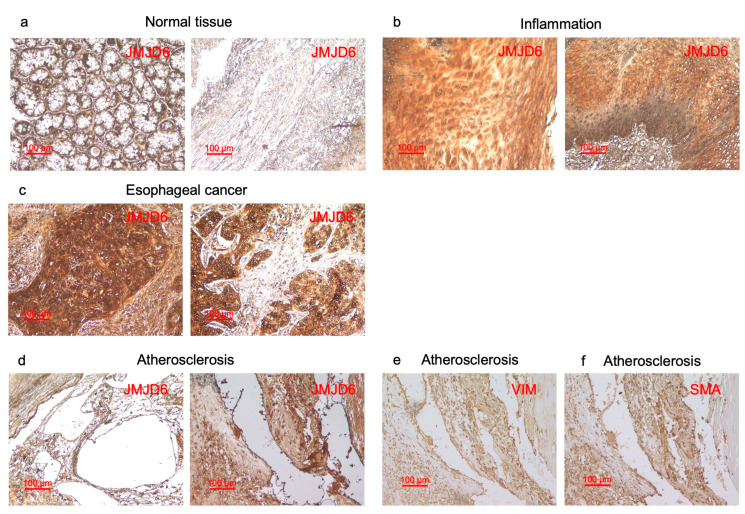
Figure 4.
Immunohistochemical staining of tissue samples against JMJD6 antibody. (a–c) This figure shows that paraffin-embedded sections of normal tissues of esophagus (a), inflamed mucosa of esophagus (b), and esophageal carcinoma (c) were stained with the antibody against human JMJD6. (d–f) This figure shows that surgically resected atherosclerotic plaques were stained using antibodies against human JMJD6 (d), vimentin (VIM) (e), and smooth muscle actin (SMA) (f). The scale bar indicating 100 µm is shown in red.

