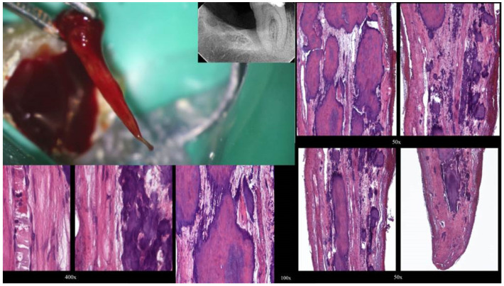
Figure 3.
Histological images of the calcified vital pulp tissue that was removed during the root canal treatment of a second mandibular molar with deep periodontal distal lesion suffering from symptomatic irreversible pulpitis. Notice the linear calcified nodules formed along the root pulp vessels (hematoxylin–eosin staining) (clinical and radiographic images are courtesy of Dr. Chaniotis Antonis, and histological images are courtesy of Prof. Domenico Ricucci).

