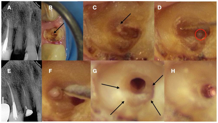
Figure 6.
(A) Preoperative radiograph of a calcified lateral incisor that suffered a crown fracture. The root canal treatment negotiation was initiated but stopped because of the non-detectable canal and misorientation. The root canal is not visible in the radiograph. (B) Clinical view of the access cavity after cleaning the debris. The arrow points to a white spot, indicating a possible canal. (C) Higher magnification clinical view. The peripherical dentine is yellow, followed by a central circular grey area that holds a white spot of accumulated debris in the white spot (black arrow). (D) A D-finder file (Mani, Japan) negotiating the calcified canal (initial catch). The red circle indicates the arrested misoriented previous access. (E) Postoperative radiograph. (F) Clinical view of the initial glide path file removing pieces of coronal restrictive dentin. (G) Clinical view of the calcified canal after the shaping procedures. Transillumination reveals the radial orientation of the dentinal tubules. The reflection of the microscope light gives the characteristic butterfly effect impression (arrows). (H) The clinical view of the gutta-percha cut deep inside the canal during post-space preparation (clinical and radiographic images courtesy of Dr. Chaniotis Antonis).

