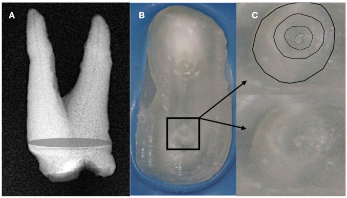
Figure 7.
(A) Radiographic image of an extracted partially calcified maxillary premolar. (B) Microscopic view of the cross-section of the calcified orifices of the extracted maxillary premolar. (C) Magnified microscope view of the area in the square. Notice the circular deposits of calcified material in the periphery reflecting different stages of tertiary, reactionary, or reparative dentine formation. Concentrating on these circular deposits, a grey area exists with a white spot of debris accumulation in the center. This white spot is indicative of the canal’s location (clinical and radiographic images courtesy of Dr. Chaniotis Antonis).

