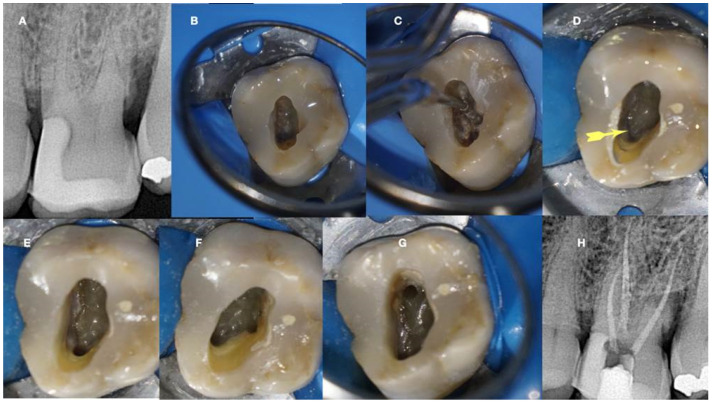
Figure 12.
Calcified maxillary molar negotiation (A). Preoperative radiograph of the maxillary molar with no detectable canals (B). A clinical view of the access cavity reveals a completely calcified chamber (C). Clinical view of the ultrasonic troughing of the pulp floor. Notice that the pulp floor color is grey (D). Clinical view of the axial wall floor junction developmental line (yellow arrow). The canal entrances in a maxillary molar always originate from this line (E–G). Clinical image of the shaped canals. Notice that the location of the canals in a calcified maxillary molar is always lying along the wall–floor junction developmental lines (H). Postoperative radiograph of the calcified maxillary molar treatment (clinical images and radiographs courtesy of Dr. Chaniotis Antonis).

