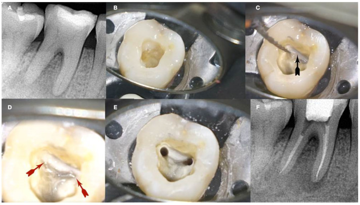
Figure 13.
Calcified mandibular molar negotiation (A). Preoperative radiograph of a calcified mandibular molar with no visible canals (B). Clinical view of the calcified chamber (C). Clinical view of the calcified tissue blocking the access to the mesial canal system. Notice the white color of the calcification. The axial walls are yellow, and the pulp floor is always grey (D). Clinical view of the pulp floor developmental lines after the removal of the white calcified tissue. Chasing the dark grey developmental lines to their termini will reveal the calcified canal entrances (E). Clinical view of the canals shaped (F). Postoperative radiograph of the calcified mandibular molar canal treatment (clinical images and radiographs courtesy of Dr. Chaniotis Antonis). Black arrow: calcified structure covering the canal orifices and the developmental pulp chamber lines, red arrows indicate the canal orifices after the removal of the white calcified structure (black arrow).

