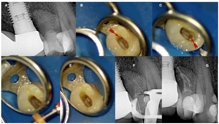
Figure 14.
Calcified maxillary premolar negotiation (A). Preoperative radiograph of a calcified maxillary premolar associated with apical periodontitis (B). Clinical view of the palatal calcified canal orifice (red arrow) (C). Clinical view of the buccal calcified canal orifice after chasing the pulp floor developmental line to its far end (red arrow) (D). Clinical view of the EDM file buckling resistance activation test negotiation (E,F). Clinical and radiographic images of the calcified canal negotiation, (G) postoperative radiograph of the calcified maxillary premolar treatment (clinical images and radiographs are courtesy of Dr. Chaniotis Antonis).

