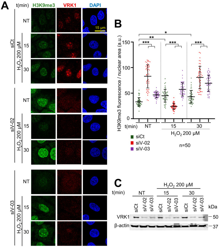
Figure 5.
VRK1 depletion facilitates the trimethylation of H3K9 in A549 lung adenocarcinoma cells exposed to hydrogen peroxide. (A) Image panels showing trimethylation of H3K9 levels stained by IF and detected using confocal microscopy. VRK1 was depleted using two siRNAs (siVRK1-02 and siVRK1-03) for 72 h. Cells were treated with 200 µM H2O2 for 15 and 30 min. (B) Quantification of H3K9me3 fluorescence per nuclear area (a.u.) of 50 cells (per condition). (C) VRK1 levels were detected by immunoblot and are shown at the bottom. β-actin was used as a loading control. Scale bar = 15 µm; * p < 0.1; **; p < 0.01; *** p < 0.001; NT: non-treated; siCt: siControl; siV-02: siVRK1-02; siV-03: siVRK1-03. Experiments were independently performed three times.

