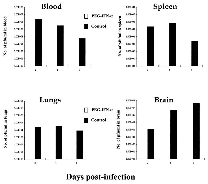
FIG. 3.
VEEV titers in blood, spleen, lung, and brain tissues of PEG IFN-α-treated and untreated BALB/c given a subcutaneous challenge with virulent VEEV. Groups of 15 mice were given 100 μl i.p. of either 4 × 10⁴ U of PEG IFN-α in 1% NMS in N-saline (□) or just 1% NMS in N-saline (■) on days −2 to +5 after infection with ca. 25 MLD (25 PFU) of VEEV in 100 μl of L-15 MM on day 0 p.i. Viral titers were calculated by standard plaque assay, incubating log dilutions of particular cell suspensions with BHK cells. Results shown are geometric mean viremias of five individual mice per time point + geometric confidence limits of 95%.

