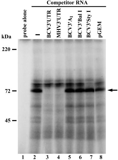
FIG. 3.
UV cross-linking of cellular proteins that bind to the BCV 3′ UTR RNA. 32P-labeled BCV3′UTR RNA probe was incubated in the absence of cell extract (lane 1) or in the presence of 30 μg of BCV-infected HCT cell extract (lanes 2 to 8). Samples were UV cross-linked for 30 min, RNase treated, heated at 95°C for 3 min, and resolved by SDS-PAGE (12% polyacrylamide). The positions of protein standards are indicated on the left. The arrow indicates the position of the p73 protein. To map the p73 protein binding site, UV cross-linking was performed in the presence of a 100-fold molar excess of coronavirus-specific unlabeled RNAs (BCV3′UTR, MHV3′UTR, BCV3′A1, BCV3′BalI, and BCV3′StyI) or 500-fold molar excess of pGEM RNA. Competitor RNAs were preincubated with lysate prior to the addition of 32P-labeled BCV3′UTR probe.

