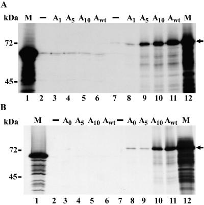
FIG. 8.
In vitro binding of PABP to coronavirus 3′ UTR RNAs. In vitro-translated luciferase or PABP was incubated with 1 μg of biotinylated BCV3′UTR RNAs containing poly(A) tails of 1, 5, 10, or 68 A residues (A) or MHV3′UTR RNAs containing poly(A) tails of 0, 5, 10, or >50 A residues (B). Immobilized streptavidin was added to recover biotinylated RNA complexes, and samples were washed to remove any unbound RNA or protein. Samples were analyzed by SDS-PAGE (8% polyacrylamide). M (lanes 1 and 12) denotes marker and corresponds to the input amount of radiolabeled luciferase (lane 1) or PABP (lane 12) in each reaction. Lanes 2 and 7 represent the level of background protein binding in the absence of biotinylated RNA for luciferase (lane 2) and PABP (lane 7). (A) Lanes 3 to 6, luciferase recovered from interaction with BCV3′UTR RNAs; lanes 8 to 11, PABP recovered from interaction with BCV3′UTR RNAs. (B) Lanes 3 to 6, luciferase recovered from interaction with MHV3′UTR RNAs; lanes 8 to 11, PABP recovered from interaction with MHV3′UTR RNAs.

