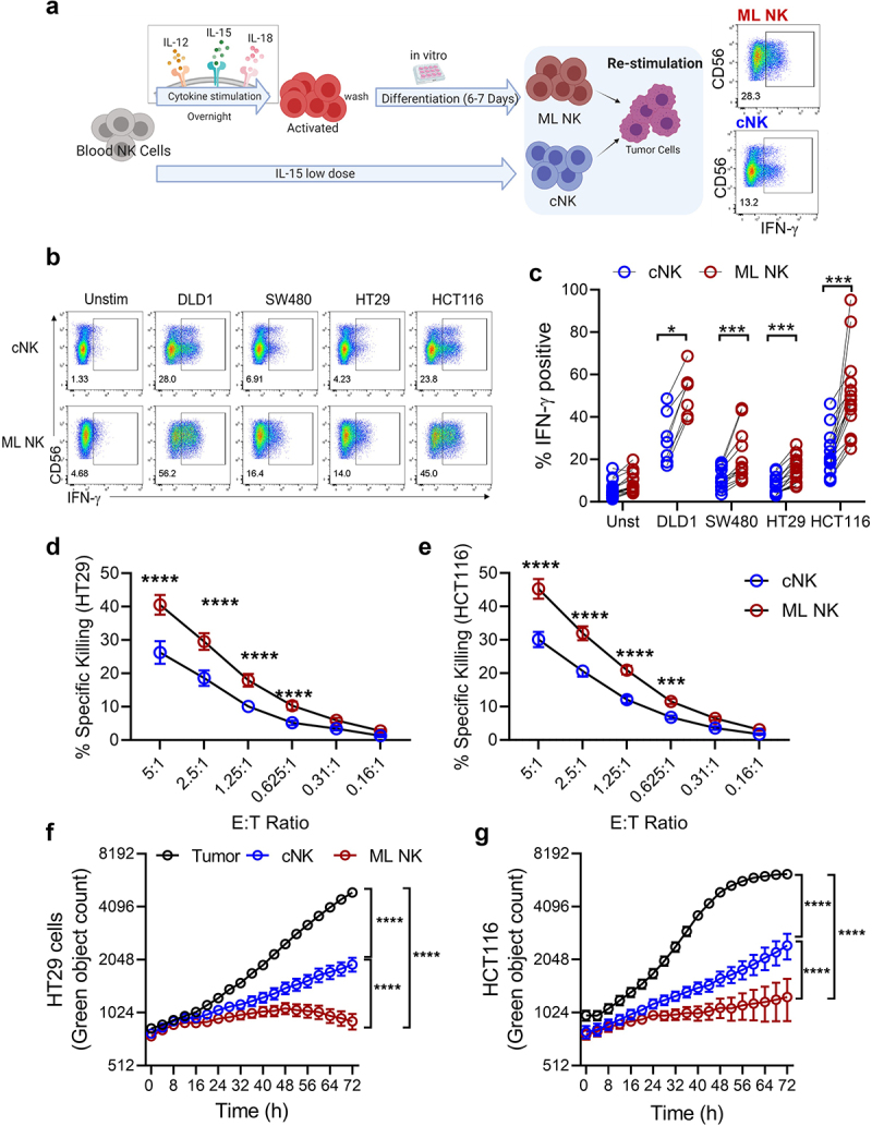
Figure 1.

ML NK cells from healthy donors exhibit enhanced functionality against colorectal cell lines. a) schema shows the generation of ML NK cells and cNK cells. b, c) IFN-γ production by cNK (b, upper) and ML NK (b, lower) cells stimulated with all four CRC cell lines. Control and ML NK cells were incubated with HT29 and HCT116 at different E:T ratios and specific lysis was assessed in a 51Cr release assay (d, e) and incucyte (f, g). For incucyte, GFP expressing HT29 and HCT116 cells were incubated with NK cells at 2.5 E:T ratio for 72 h. F and G show one representative example from 3 different donors in 3 independent experiments. n = 7–12 in C, n = 3 in D and E. Two-way ANOVA with Sidak post test. *p < 0.05, ***p < 0.001, ****p < 0.0001.
