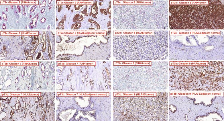
Fig. 1. HLA-E expression in PCa.
IHC analysis of PCa or adjacent normal tissue for expression of HLA-E, PAP, and PIN4. TMAs were prepared and processed as described in Materials and Methods. Representative IHC of PCa tumor tissue and adjacent normal tissue are shown for Gleason 7, 8, and 9 tumors stained for HLA-E using a polyclonal antiserum. Pathological tumor (pT) stages of PCa are indicated with pT2c representing localized bilateral PCa, whereas pT3b represents locally advanced PCa. Staining for PAP and PIN4 is additionally shown for the tumor tissue. The results for all TMAs are summarized in data file S1.

