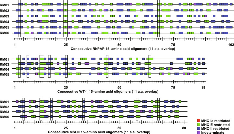
Fig. 3. Epitope specificity and MHC restriction of TAA-targeting CD8+ T cells.
CD8+ T cell responses in PBMC of RMs 1 to 6 (Fig. 2) were measured by ICS for IFNγ and TNFα to individual overlapping consecutive 15–amino acid oligomer peptides that span the indicated proteins. Assay limit of detection was determined as described previously (51), with 0.05% after background subtraction being the minimum threshold. Above background responses (IFNγ and/or TNFα) are shown by a square along the length of each protein with the peptide numbers shown below. Results of blocking experiments are indicated by the color of the square: Peptides restricted by MHC-II (blue) were blocked with class II-associated invariant chain peptide (CLIP) and anti–human leukocyte antigen-DR (HLA-DR) antibody, whereas MHC-E–restricted peptides (green) were blocked with VL9 peptide and pan–MHC-I antibody. Indeterminate responses are shown in purple. Supertopes, i.e., epitopes recognized in all RMs (including Fig. 5), are boxed in. Overall analysis of 15–amino acid oligomer peptide responses is shown in data file S3. a.a., amino acid.

