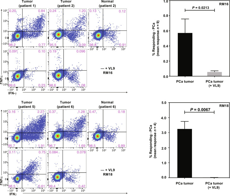
Fig. 8. Recognition of primary PCa cells by MHC-E–restricted, PAP-specific CD8+ T cells.
Left: ICS for IFNγ and TNFα production by CD8+ T cells from two 68-1 RhCMV/RhPAP-immunized RM after coincubation with PCa cell suspensions in the presence or absence of VL9 peptide. In addition, we inhibited MHC-II presentation by adding anti-DR antibody and CLIP peptide in all stimulations. Right: Summary of results from the indicated number of primary PCa samples showing the average frequency of CD69 and IFNγ and/or TNFα-positive CD8+ T cells from 68-1 RhCMV/RhPAP-immunized RM in the absence or presence of VL9 peptide after background subtraction. Individual results are shown in data file S3. Statistical significance was calculated using paired t test.

