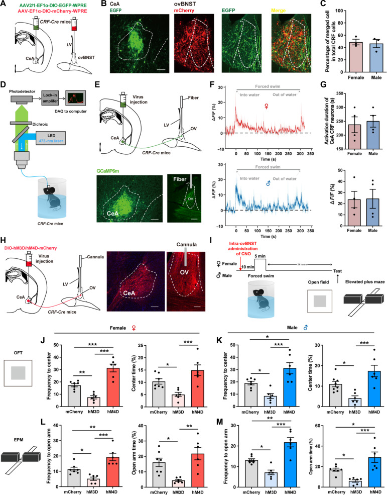
Fig. 2. CeA CRF projections to the ovBNST mediate stress and anxiety in both sexes.
(A) Schematic diagram of dual viral injections into the ovBNST and CeA of CRF-Cre mice for anterograde monosynaptic tracing. (B) A part of mCherry+ CRF neurons in the ovBNST were labeled with EGFP. Scale bar, 200 μm. (C) Quantification of the percentage of merged cell in total CRF cells. (D) Experimental setup for fiber photometry. (E) Top: Virus injection and fiber configuration. Bottom: Representative image showing expression of GCaMP6m in CeA and ovBNST. Scale bars, 200 μm. (F) Example trace of calcium signals for forced swimming application in female (top) and male (bottom) mice. (G) Quantification of forced swimming-induced activation duration (top) and peak ΔF/F (%) (bottom) for female and male mice. (H) Left: Virus injection and cannula configuration. Right: Representative image showing expression of hM3D/hM4D-mCherry in CeA and terminals in ovBNST. Scale bars, 200 μm. (I) Experimental timeline. (J and K) Quantification of frequency to center zone and percentage time spent in the center zone of OFT in female (J) and male (K) mice [*P < 0.05, **P < 0.01, and ***P < 0.001, one-way analysis of variance (ANOVA) and post hoc test]. (L and M) Quantification of frequency to open arm and percentage time spent in the open arm of EPM in female (L) and male (M) mice (*P < 0.05, **P < 0.01, and ***P < 0.001, one-way ANOVA and post hoc test). LED, light-emitting diode; DAQ, data acquisition; OV, oval nucleus; CeA, central amygdala; LV lateral ventricle.

