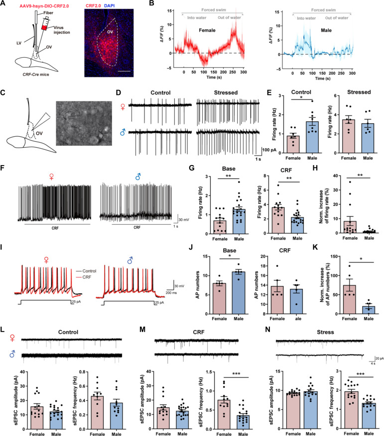
Fig. 3. CRF release is sexual different in ovBNST during stress and drives hyperexcitation of ovBNST CRF neurons in female mice in vitro.
(A) Left: Virus injection. Right: Representative image showing expression of CRF2.0 in ovBNST. Scale bar, 200 μm. (B) Example trace of CRF release in ovBNST during forced swimming stress in female (left) and male (right) mice. (C) Schematic of whole-cell patch-clamp recordings of ovBNST neurons. (D) Representative firing rate of ovBNST CRF neurons before and after stress in female (up) and male (down) mice. (E) Quantification of the firing rate of ovBNST CRF neurons before (left) and after (right) stress (*P < 0.05, Student’s t test). (F) Representative firing frequency of ovBNST neurons after perfusion of CRF in female (left) and male (right) mice. (G) Quantification of the firing rate before (left) and after (right) CRF perfusion in female and male mice (**P < 0.01, Student’s t test). (H) Quantification of the normalized increase of firing rates after CRF perfusion in female and male mice (**P < 0.01, Student’s t test). (I) Representative evoked firing activity was recorded before and after perfusion of CRF in female (top) and male (bottom) mice. (J) Quantification of the evoked AP numbers before (left) and after (right) CRF perfusion in female and male mice (*P < 0.05, Student’s t test). (K) Quantification of the normalized increase of evoked AP numbers after CRF perfusion in female and male mice (*P < 0.05, Student’s t test). (L to N) Example sEPSC traces of ovBNST CRF neurons and quantitative analysis of sEPSC amplitude and frequency in control (L), after CRF perfusion (M) and after stress (N) in female and male mice (***P < 0.001, Student’s t test). DAPI, 4′,6-diamidino-2-phenylindole.

