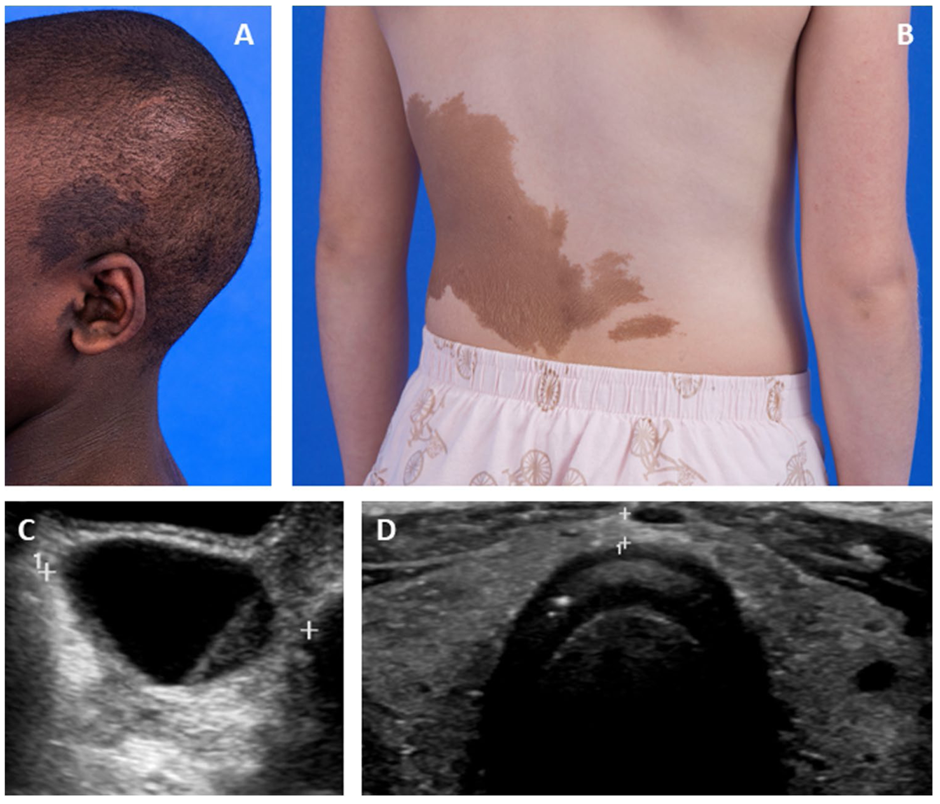
Fig. 1.

Extraskeletal features of McCune-Albright syndrome. A Photograph showing typical hyperpigmented macules overlying a patient’s skull and ear. B Photograph showing typical hyperpigmented macules on a patient’s trunk. C Pelvic ultrasound image showing a typical ovarian cyst associated with precocious puberty. D Thyroid ultrasound image showing diffuse heterogeneity of both lobes
