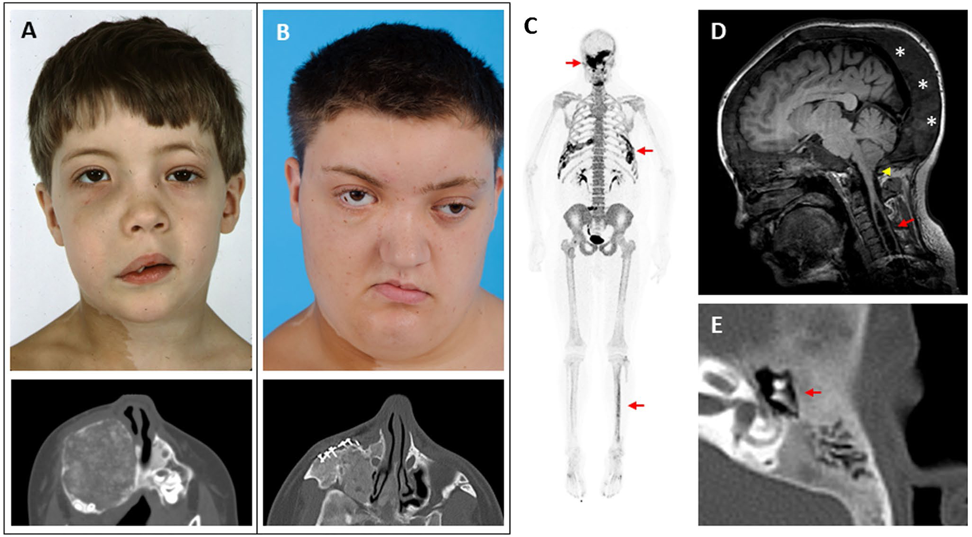
Fig. 2.

Clinical images of craniofacial fibrous dysplasia (FD). The left-hand panels show images from the same patient at ages 6 (A) and 18 years (B). Note the progression in facial asymmetry, including expansion of the right-sided face and orbital dystopia. The lower panels show axial computed tomography views of his right maxilla, including surgical implants from a reconstructive procedure performed at age 7. C 18F-NaF PET/CT scan performed in this patient at age 25 years. Areas of increased tracer uptake correspond to FD lesions in his skull, ribs, and tibia (red arrows). D Brain MRI from a 17-year-old girl with craniofacial FD and headaches. The cerebellar tonsils extend below the foramen magnum, consistent with a Chiari I malformation (yellow arrowhead). An associated syrinx has developed in the spinal cavity (red arrow). Note the diffuse expansion of the posterior cranium involved with FD (asterisks). E Computed tomography scan of the temporal bone in a 13-year-old patient with mild conductive hearing loss. The etpitympanum is diffusely involved with FD (red arrows), surrounding the ossicular chain
