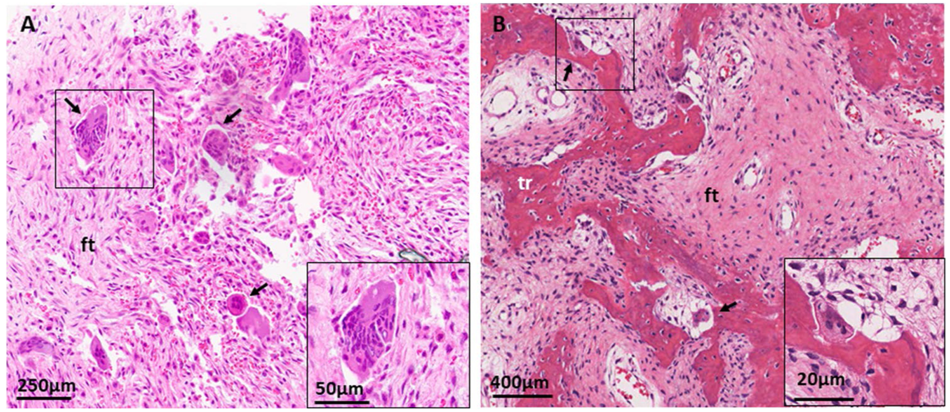
Fig. 3.

Histologic features of craniofacial fibrous dysplasia. A H&E section from a sphenoid bone shows diffuse fibrous tissue (ft) with prominent, irregularly situated osteoclasts (black arrows). B H&E section from a maxilla shows discontinuous, curvilinear trabeculae (tr) on a background on fibrous tissue (ft). Again noted are prominent osteoclasts (black arrows)
