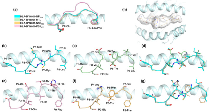
Figure 4.

Crystal structures of the HLA‐B*18:01 binding cleft, presenting four immunogenic influenza peptides. (a) Structural overlay of the peptide backbone and anchor positions (cartoon) for NP219 (cyan), M15 (green), NS2111 (yellow) and PB1117 (pink), within the binding cleft of HLA‐B*18:01 (light cyan). Side view of the HLA‐B*18:01 binding cleft presenting NP219 (b), M15 (c) and their overlay (d). Side view of the HLA‐B*18:01 binding cleft presenting PB1117 (e), NS2111 and their overlay (f). (b–g) Peptide residue side chains are represented in stick. (h) Top‐down view of NS2111 is represented as a transparent surface (white) within the binding cleft of HLA‐B*18:01 (light cyan cartoon).
